
Posterior Diagnostic
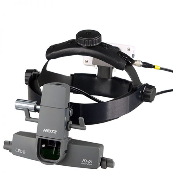
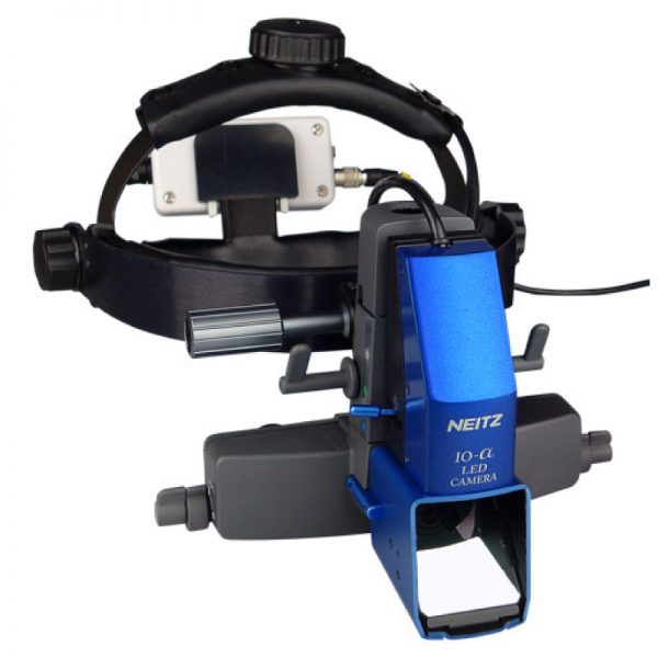
Binocular Indirect- IO-α LED
- An LED with the color of incandescent is used
- Clear illumination without filamant shadow
- Stereoscopic observation even for small pupils by moving the knobs of the illumination and observation systems separately
- Large effective visual field for observation of flat pupils
- 17 hours of continuous illumination with the energy-efficient LED
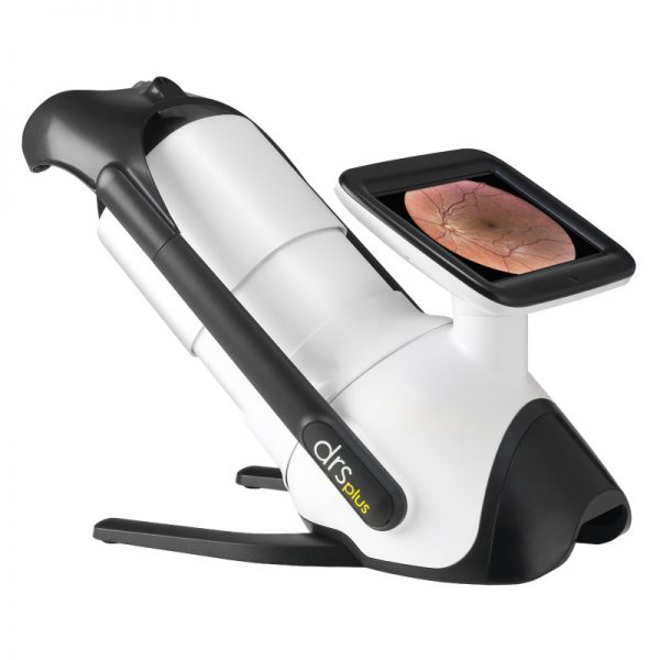
DRSplus
- TrueColor Confocal Technology
- Multiple imaging modalities including Red-free, external eye and stereo view imaging
- 2.5 mm minimum pupil size
- Fast, easy and fully automated operations
- Mosaic function which creates retinal panoramic views up to 80°
- Remote Viewer that allows for reviewing from devices on the same local area network
- Remote Exam feature enables executing an exam from a distance
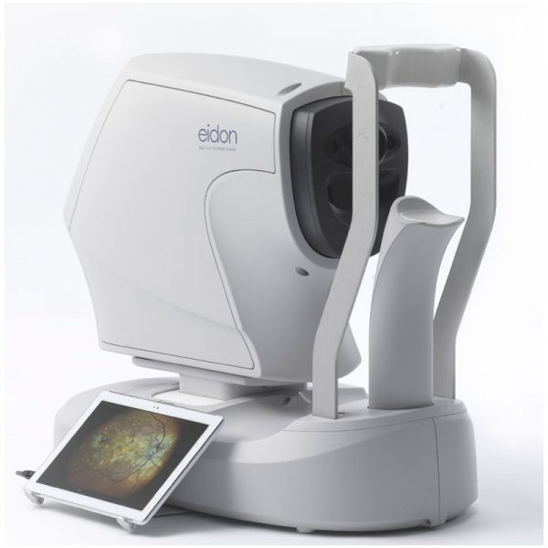
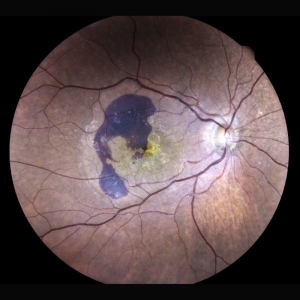
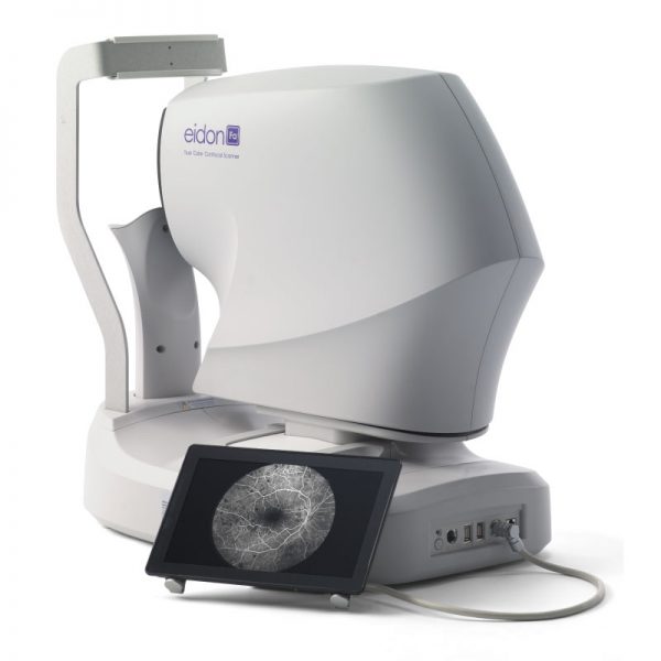
Eidon
- Multiple imaging modalities including TrueColor, blue, red and Red-Free and infrared confocal images
- Widefield, ultra-high-resolution imaging
- Capability to image through cataract and media opacities
- Dilation-free operation (minimum pupil 2.5 mm)
- Flexibility of fully automated and fully manual mode
- All-in-one compact design, no additional PC required
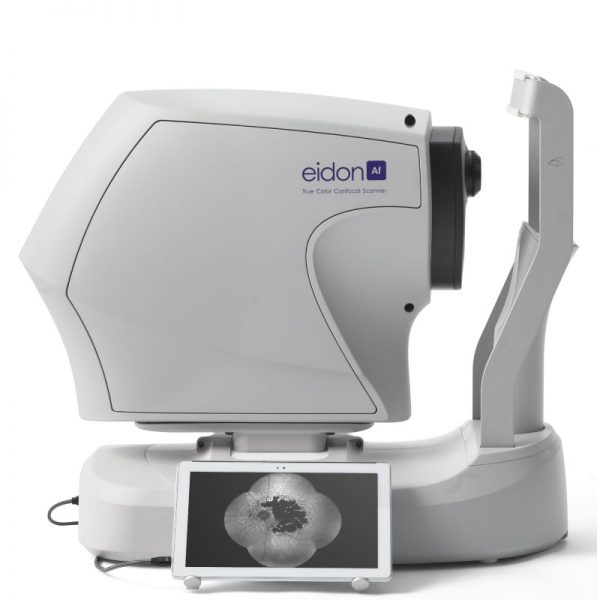
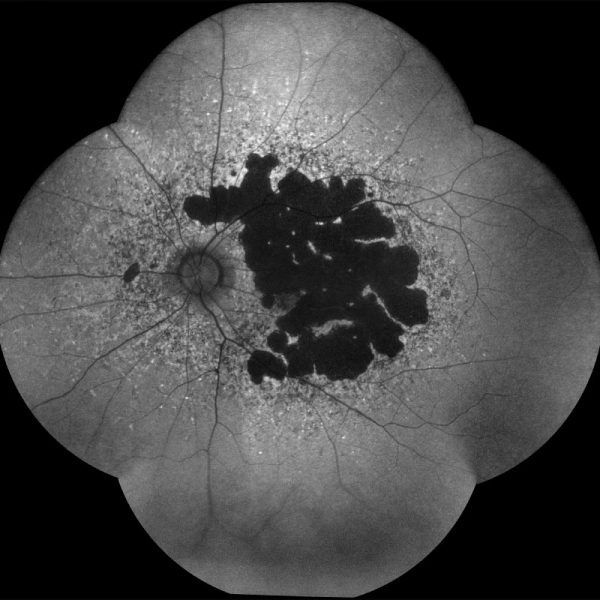
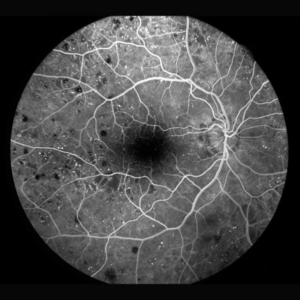
Eidon FA
- Fundus Imaging system with Color,AF, FA pictures
- Each picture has 90 degree angle or 120 degree with UWFL
- Includes all the features and functionalities of EIDON technology
- Complete suite of Fundus imaging capabilities
- High resolution Fluorescein Angiography images
- High-resolution and dynamic Fluorescein Angiography video view
- TrueColor confocal technology for high-quality, accurate imaging
- Fully automatic, easy-to-use and requires minimal staff training
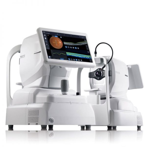
HOCT-1F
- High-speed and high-quality scanning (68,000 A-scans per second)
- 3D rendering of retina and macula thickness/separation
- Combines OCT, fundus camera, angiography (HOCT-1F/FA), Topography,Biometry and PC all in one device
- Web browser allows you to analyse data independently from the machine with no need to install specialist software
- Detailed reports showing pathological structure and important data in an easy-to-read format that can be viewed on-screen or printed
- Anterior Segment Module allows measurement of corneal thickness, angle and 3D imaging.
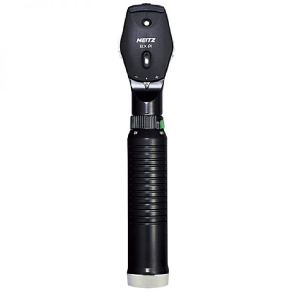
Ophthalmoscope – BXα-RC
- The correction lens from -36 D to +35 D at the interval of 1 D to focus accurately
- Unique polarizing filter for observation of natural fundus images
- Neitz original optical systems for minimal corneal reflex
- Dust shutter to prevent foreign matter
- Rechargeable battery powered. No need for battery replacement
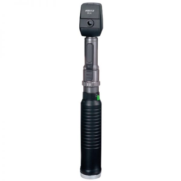
Streak Retinoscope-RX-RC
- Use bulbs with precisely processed filaments of 0.05mm diameter, which create one of the sharpest streaks of light in the industry
- Helps the accurate diagnosis of the astigmatic axis
- The beam can be turned 360 degrees
- The anti-reflection filter provides a brighter and wider field of view - Rechargeable battery powered, no need of battery replacement


